Cutting-edge tech captures images inside human heart arteries, revolutionizing medical science
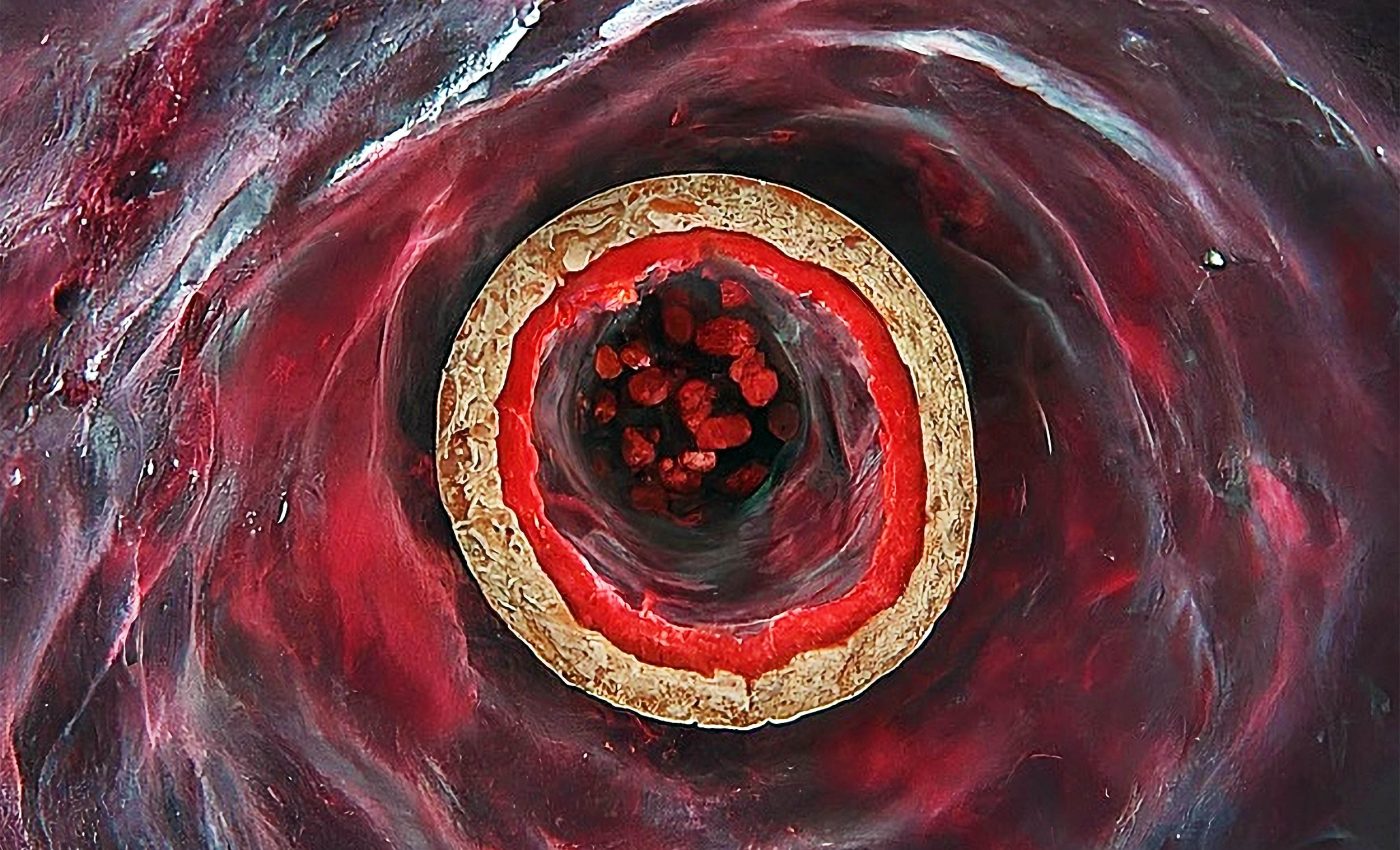
Heart doctors are capturing images showing the inside of heart arteries, helping them watch plaque respond to powerful cholesterol drugs in real-time.
In these studies, PCSK9 inhibitors, drugs that block a cholesterol controlling protein, made plaques smaller and more stable within about one year of treatment.

The pictures come from international trials that scanned the same artery segments in hundreds of patients before and after therapy.
Teams at major hospitals in Europe and North America combined their data to see how these drugs change arteries deep inside the body.
Huge impact of artery imaging
For many years, doctors tracked coronary atherosclerosis, a slow buildup of fatty plaque in heart arteries, with angiograms that only outlined the vessel channel.
Those pictures showed where arteries narrowed but revealed almost nothing about the hidden material inside the vessel wall.
A recent review pulled together studies that used invasive imaging to watch how PCSK9 based treatment changes coronary plaque in living patients.
The work was led by Massimiliano Ruscica, PharmD, PhD, at the University of Milan, whose research centers on PCSK9, atherosclerosis, and lipid lowering therapies.
Cholesterol and plaque
For decades, treatment focused on lowering low density lipoprotein cholesterol, a blood fat that drives plaque growth, and counting heart attacks and strokes.
Statins already reduced those events, yet many patients still had trouble even when their lab numbers looked fine.
PCSK9 inhibitors keep more LDL receptors active in the liver, so the body clears extra LDL particles from the bloodstream. As a result, they can push LDL cholesterol far below levels that most people reach with statins alone.
The FOURIER outcomes trial showed that adding evolocumab to statins dropped LDL to about 30 milligrams per deciliter and reduced major cardiovascular events in high risk patients.
That result raised a sharp question about whether the benefit came only from changing blood numbers or also from reshaping plaque inside the artery wall.
Answering that question meant looking directly at the plaques instead of guessing from cholesterol tests. New invasive imaging tools made it possible to watch plaque in the same artery segment change over time.
Intravascular cameras
New tools such as intravascular ultrasound, a catheter based ultrasound that images artery walls from inside, let cardiologists measure plaque volume rather than just the open channel.
The same catheter run can be repeated months later, giving matching before and after pictures of the very same vessel segment.
Light based optical coherence tomography, a high resolution imaging method using reflected light, can see the thin fibrous layer that covers the soft core of a plaque.
It can measure how thick that cap is, which matters because very thin caps are more likely to tear and release clot forming material.
Near-infrared spectroscopy (NIS) uses infrared light absorption patterns to map how much oily lipid sits inside plaque cores.
When combined with ultrasound or optical imaging, it helps identify plaques that are especially rich in lipid and therefore more dangerous.
Taken together, these invasive cameras give several complementary views of the same disease. One measures size, another shows structure, and a third highlights the chemical makeup inside the plaque.
PCSK9 and dangerous plaques
In the GLAGOV trial, nearly one thousand statin treated patients had coronary plaque measured from inside the artery using intravascular ultrasound.
Adding evolocumab produced a clear drop in the percentage of the artery wall filled with plaque, and more than half of the patients showed actual regression of plaque volume.
A later study called HUYGENS used optical coherence tomography and ultrasound in patients who had a type of heart attack called non ST elevation myocardial infarction.
Evolocumab on top of statins made the protective cap thicker and reduced the arc of lipid inside plaques compared with placebo.
The PACMAN-AMI trial tested the PCSK9 blocker alirocumab in three hundred patients after an acute myocardial infarction, a sudden blockage of a heart artery that kills muscle.
Serial imaging showed that alirocumab plus high intensity statin therapy shrank plaque burden, reduced lipid rich core, and increased cap thickness in arteries that had not caused the original heart attack.
In people born with very high cholesterol, a condition called familial hypercholesterolemia, an inherited disorder that causes lifelong high LDL, the ARCHITECT trial followed more than one hundred patients on alirocumab and strong statins.
Over roughly a year and a half, overall plaque burden in the coronary tree fell, and the unstable fibrofatty and necrotic core portion shrank while more stable fibrous and calcified tissue increased.
Why the plaque changes matter
Cardiologists watch features such as a large lipid core, a very thin fibrous cap, a scar-like tissue layer over plaque, and high inflammation because they signal a plaque that can rupture.
These risky plaques may sit in arteries that look only mildly narrowed on standard angiograms, yet they are often the ones that break and trigger clots.
Imaging trials now suggest that PCSK9 inhibitors shift plaques away from this high risk pattern toward a quieter, more stable state.
In these studies, plaques tended to become smaller, more fibrous, less packed with free cholesterol, and better covered by thicker caps that are less likely to split.
These shifts in plaque features match what doctors expect when LDL is driven very low for long periods. The imaging results link changes in blood tests to physical changes in plaque, which makes the clinical benefits of intensive treatment easier to understand.
Because the imaging work follows the same patients over time, it connects LDL reductions with direct evidence of plaque healing in those arteries.
That picture helps explain why treating aggressively, sometimes within days after a heart attack, can change long term risk.
Lessons from artery imaging
These studies encourage a move from caring only about how narrow an artery looks to caring about what kind of plaque is present.
A modest narrowing made mostly of soft lipid with a thin cap can be more dangerous than a tighter narrowing built from tough fibrous and calcified tissue.
Researchers are testing whether noninvasive scans guided by plaque features, such as low density regions or changes in surrounding fat, can flag people who most need aggressive LDL lowering.
If that strategy proves reliable, doctors might one day tailor PCSK9 therapy not just to cholesterol numbers, but also to the hidden biology of each patient’s plaque.
At the same time, PCSK9 inhibitors remain costly and are usually reserved for people at very high risk who do not reach LDL targets with other treatments.
Imaging could help make those decisions more precise by identifying patients whose plaque still looks dangerous despite apparently good standard therapy.
Ongoing trials are exploring how early and how intensively to use these drugs after a heart attack, and whether imaging endpoints should help steer future guidelines.
As more data emerge, artery cameras may play a growing role in showing when plaque has truly calmed and when more treatment is still needed.
The study is published in Current Atherosclerosis Reports.
—–
Like what you read? Subscribe to our newsletter for engaging articles, exclusive content, and the latest updates.
Check us out on EarthSnap, a free app brought to you by Eric Ralls and Earth.com.
—–


