520-million-year-old fossil with brain and guts intact is called an 'archeological miracle'
Animal life took off quickly during the Cambrian Period, and every so often, a tiny fossil discovery reminds us how quickly complexity took shape.
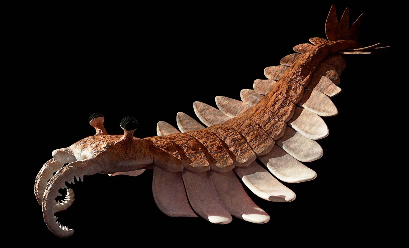
A newly described fossil of an arthropod named Youti yuanshi was found preserved with its soft tissues intact after roughly 520 million years.

A recent analysis of this early life form offers an unusually clear look at the insides of one of the earliest relatives of insects, crabs, spiders, and their kin. It is small, ancient, and surprisingly well organized internally.
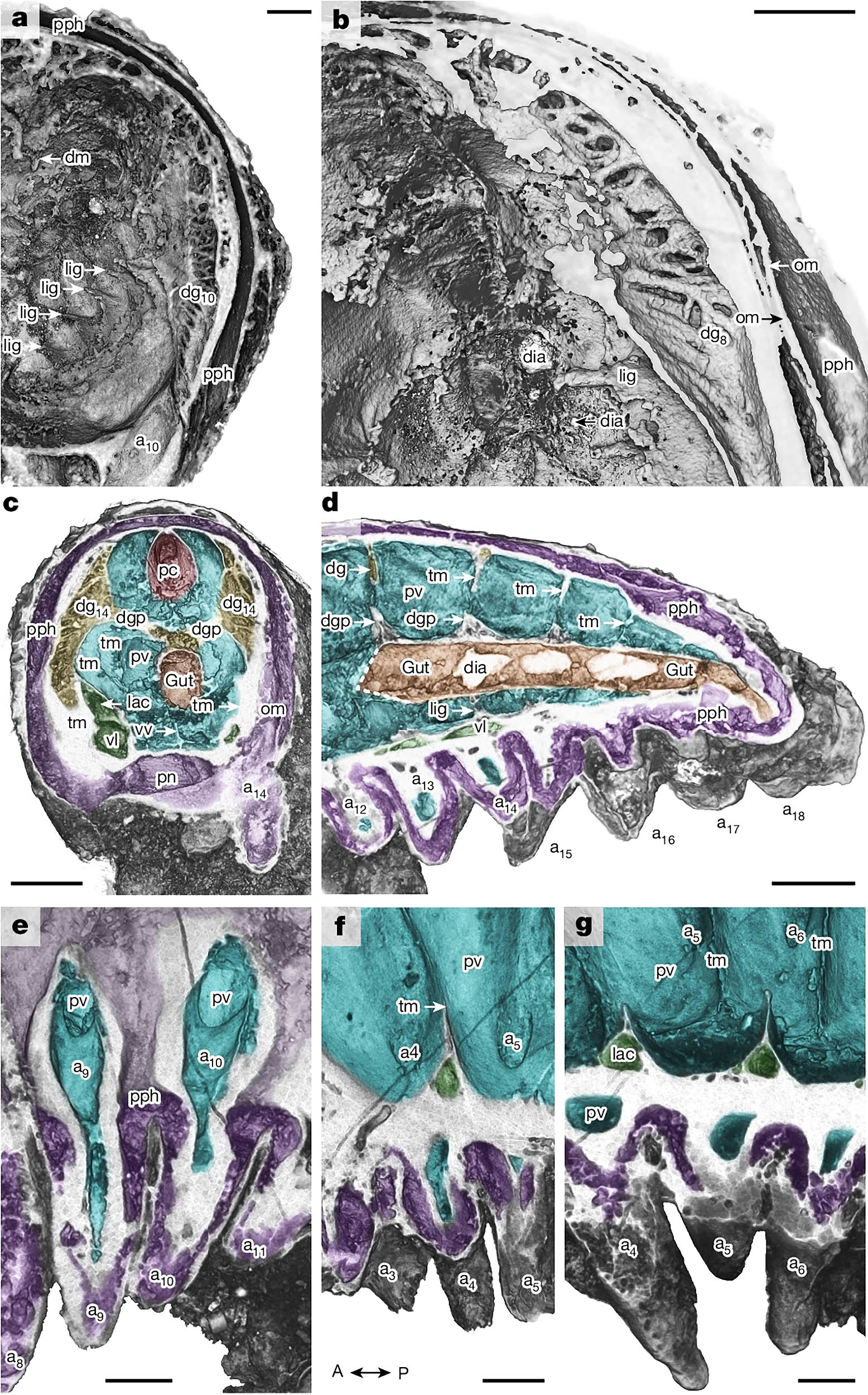
The fossil captures internal anatomy in three dimensions rather than as a flattened smear.
Researchers used powerful X-ray imaging to virtually examine the body and map structures that usually vanish after death. This preservation gives scientists a way to trace how early animal body plans came together.
Discovering Youti yuanshi
The fossil comes from Yunnan, China, a region known for Cambrian rocks that often capture delicate details.
The team named the larva Youti yuanshi in a nod to its juvenile stage and its role in understanding early arthropods.
When it was alive 520 million years ago, animal groups were branching out into diverse forms and lifestyles.
Youti measures about 0.15 inches (3.8 millimeters) long. The body is divided into 20 segments, each carrying a pair of soft legs known as lobopods.
These limbs lack hard joints, reflecting an early stage in the lineage that would later give rise to the fully jointed, exoskeleton-covered limbs seen in modern arthropods.
Why Youti yuanshi stands out
Here, three-dimensional depth is preserved, letting researchers separate and identify organs that occupied different layers of the body.
“When I used to daydream about the one fossil I’d most like to discover, I’d always be thinking of an arthropod larva, because developmental data are just so central to understanding their evolution,” enthused lead researcher Dr. Martin Smith of Durham University.
Boundaries between membranes, cavities, and tubes remain visible, like a miniature anatomical map from deep time.
That clarity matters because it allows scientists to match positions and connections among organs, then compare those patterns with living arthropods and their close relatives.
When structures line up across species, they mark shared features that date back to common ancestors.
Peering inside the head
The head layout is advanced for a larval stage. The mouth sits on the underside, and a circumoral (around-the-mouth) nerve ring encircles it. Distinct brain regions lie just behind.
This arrangement creates clear landmarks: a ring of nerves that controls feeding and sensing, and brain compartments that echo the basic architecture seen in living arthropods.
When positions and connections match, researchers can trace how features shifted or specialized over evolutionary time without losing track of their original identity.
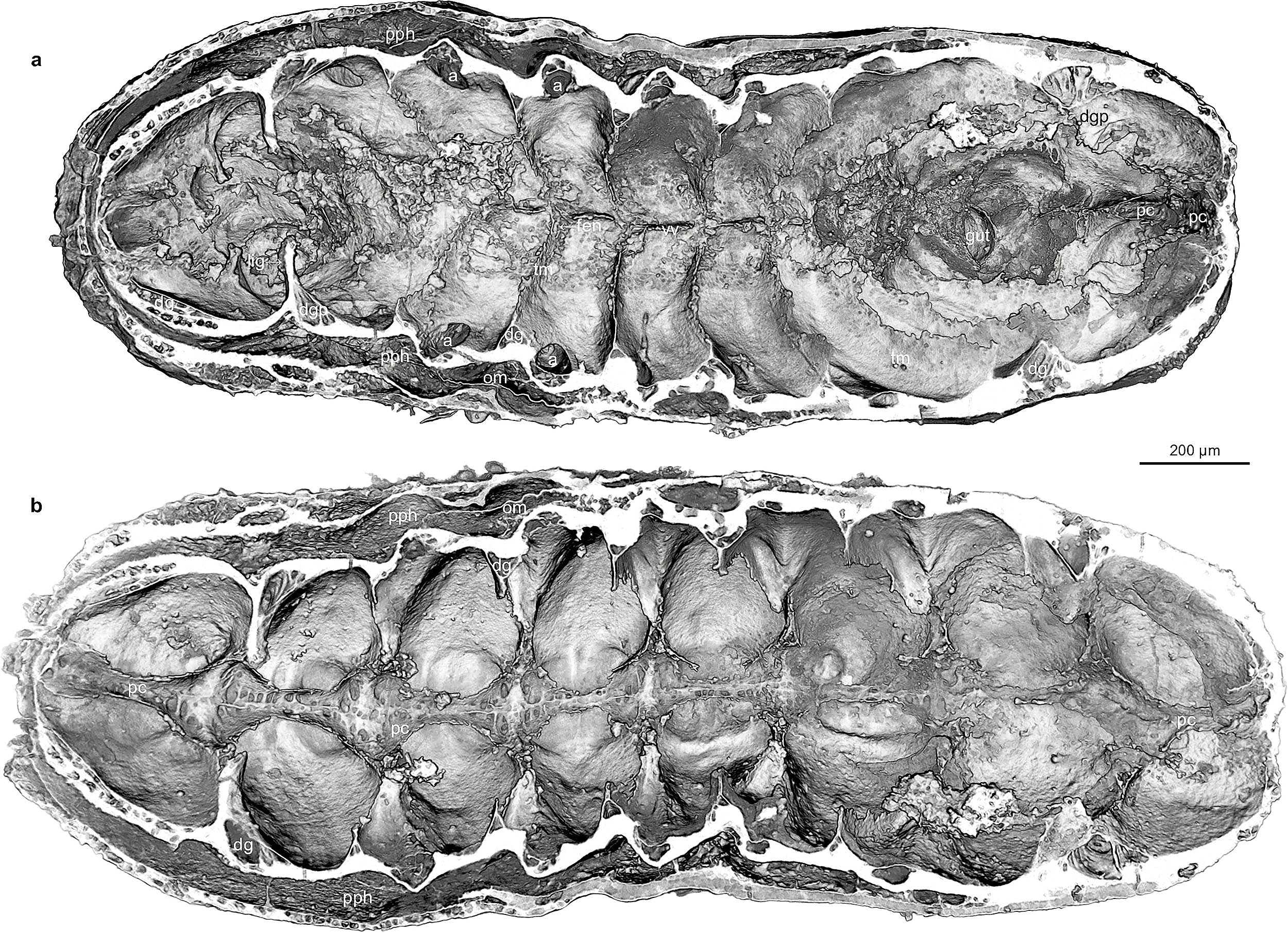
Well-defined body segments
A central digestive tract runs down the middle of the body. Along its sides, paired midgut glands help process food by increasing the surface area for digestion.
Their repeated, segmental pattern fits the idea that early arthropods modified a basic series of units – segments and their associated organs – into more specialized roles.
Circulation shows order as well. Arthropods do not have red blood like humans; their fluid, called hemolymph, moves through open spaces and channels.
In Youti yuanshi, a broad pericardial sinus sits along the top of the body, consistent with a heart-related structure, while additional sinuses run along the sides and underside, forming loops that reach the mouth region and extend into the limbs.
The layout points to a patterned system for routing fluid to tissues that need it, earlier in the group’s history than many expected.
Dissecting Youti yuanshi
The team distinguished nerves, membranes, and fluid channels by reading the X-ray slices like a stack of pages.
Each structure has telltale signs: position, shape, and the way it connects to neighbors. A nerve ring must encircle the mouth opening and link to nerve cords running into the head and trunk.
A pericardial sinus must sit above the gut and show consistent relationships to nearby membranes and limb bases across slices.
When all those clues align, the interpretation is supported.
“It’s always interesting to see what’s inside a sample using 3D imaging, but in this incredible tiny larva, natural fossilization has achieved almost perfect preservation,” noted study co-author Dr. Katherine Dobson of the University of Strathclyde.
This approach is more than a technical advance; it turns a tiny fossil into a testable set of anatomical claims for how the body was arranged.
Each claim ties to the same logic biologists use when they map body plans in living animals: where is it, what does it touch, and how does it repeat or vary along the body?
Why this ancient larva matters
Development provides a timeline for when features first appear. Early life stages often reveal the core blueprint before adult specialization takes over.
If a larva already shows a subdivided brain, a nerve ring coordinating feeding, digestive side pockets, and organized circulation, then those systems belong to the earliest set of features from which later arthropods built more elaborate bodies.
That idea fits the broader Cambrian story. Evolution did not add complexity in one sudden step.
It layered new functions onto a segmented framework, then hardened exteriors, added joints, enhanced senses, and refined head parts.
“Larvae are so tiny and fragile, the chances of finding one fossilized are practically zero – or so I thought!” Dr. Smith continued.
“I already knew that this simple worm-like fossil was something special, but when I saw the amazing structures preserved under its skin, my jaw just dropped – how could these intricate features have avoided decay and still be here to see half a billion years later?”
This combination of modularity and early specialization sets the stage for the diversity of adult designs.
Understanding Earth’s earliest life
These methods apply beyond this single species. With three-dimensional preservation and non-destructive imaging, researchers can test how early body systems were arranged, compare them to living forms, and link fossil snapshots to modern anatomy.
Early arthropods did not wait to get organized. Even at a tiny size and a juvenile stage, Youti yuanshi shows a coordinated head, a mapped-out gut with paired glands, and a structured circulatory network.
Those features appear alongside the classic segmented trunk and repeated limbs.
From that small start, the lineage had what it needed to expand across oceans, land, and air – and to keep experimenting with body plans for hundreds of millions of years.
The full study was published in the journal Nature.
—–
Like what you read? Subscribe to our newsletter for engaging articles, exclusive content, and the latest updates.
Check us out on EarthSnap, a free app brought to you by Eric Ralls and Earth.com.
—–


