Study debunks 100-year-old understanding of what brain cells look like, disproving what we've been taught
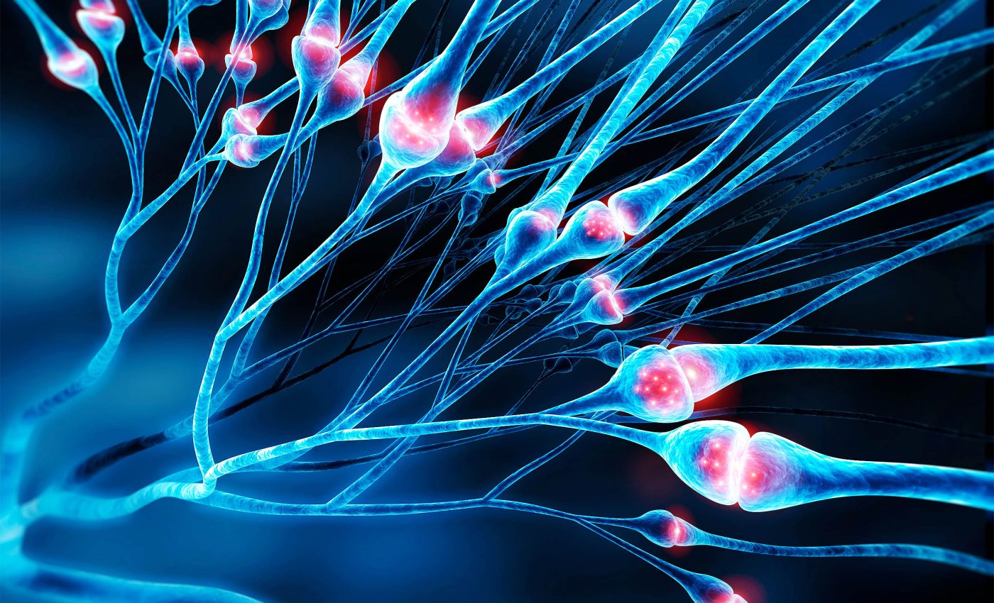
For decades, biology textbooks have told us that axons – the threadlike extensions of nerve cells – are smooth, cylindrical tubes. These axons are responsible for ferrying messages between neurons at lightning speed, playing a critical role in everything from moving your fingers to forming memories.
But new research suggests those tidy textbook images may be due for an update.

Using cutting-edge imaging methods, scientists discovered that axons don’t quite look like straight cables. Instead, they often appear as strings of tiny bubbles – like pearls on a string.
This surprising twist in what we thought was settled biology could reshape our understanding of how the brain’s wiring really works.
Axons aren’t just tubes, but pearls
What makes axons so important is their ability to rapidly transmit electrical signals, known as action potentials. These are brief bursts of electricity that allow neurons to communicate across long distances.
And speed matters – wider axons, like wider roads, help messages move faster. That’s why the shape of axons has long been thought to be smooth and uniform.
But researchers noticed something odd: using a technique called high-pressure freezing electron microscopy – which freezes brain cells in their natural shape – they didn’t see smooth tubes. They saw repeated bulges and constrictions, giving the axons a bead-like look.
These bulges are now referred to as “non-synaptic varicosities.” They aren’t just random swellings or signs of disease. They appear in healthy neurons and could actually influence how quickly messages travel through the brain.
An internal skeleton with flex
So what’s causing the pearling? Inside every axon is a structural framework called the membrane periodic skeleton, or MPS.
Think of it like a flexible scaffold made of proteins such as actin and spectrin, arranged in a repeating pattern under the membrane. This skeleton gives the axon strength and elasticity.

But here’s the catch: it’s not tightly glued to the axon’s outer membrane. That means the axon surface can wobble and bulge while the internal skeleton holds everything together.
When physical forces – like tension or pressure – change, those bulges can form naturally. It’s a well-known physical phenomenon called “pearling instability,” seen in materials ranging from soap bubbles to sausages.
How to see axon pearling structure
The research team, based at Johns Hopkins, used a freezing technique that avoids the common problem of cell distortion.
“To see nanoscale structures with standard electron microscopy, we fix and dehydrate the tissues, but freezing them retains their shape – similar to freezing a grape rather than dehydrating it into a raisin,” said Shigeki Watanabe, Ph.D., associate professor of cell biology and neuroscience at the Johns Hopkins University School of Medicine.
They looked at three types of mouse neurons: lab-grown cells, adult mouse brain tissue, and embryonic mouse neurons. All of them showed the same bubbly shape in their axons – in tens of thousands of images.
Signals, speed, and sugar
The team didn’t stop with just images. They wanted to understand whether these pearl-like shapes had any functional effects on how neurons work.
Working with theoretical biophysicist Padmini Rangamani, Ph.D., from the University of California San Diego School of Medicine, they turned to mathematical models to test different conditions.
Changing the environment around the axons – like adjusting sugar concentration or membrane tension – altered the size of the pearls.
Adding sugar made the pearls smaller, while relaxing the membrane tension had the same effect. It seems that both physical properties and the cellular environment influence how much pearling happens.
In another test, they removed cholesterol from the axon membranes, making them softer and more fluid. When they did this, there was less pearling and a noticeable drop in the axon’s ability to carry signals.
“A wider space in the axons allows ions [chemical particles] to pass through more quickly and avoid traffic jams,” said Watanabe.
Stimulating with electricity
To push the idea further, the scientists applied high-frequency electrical stimulation to the mouse neurons. This made the pearl-like segments swell – on average, they grew 8% longer and 17% wider.
These changes lasted at least 30 minutes and boosted the speed of electrical signaling. But if the membrane was cholesterol-depleted, the pearls didn’t swell, and the signaling speed didn’t improve.
That indicates a tight relationship between axon shape, membrane composition, and signal speed. It’s not just structural – it’s functional.

Scientists have known that axon beading can happen in dying cells or in neurodegenerative diseases like Parkinson’s. But seeing it regularly in healthy cells turns old assumptions on their heads.
These axon pearl shapes may not be signs of damage but adaptive features that help neurons function more efficiently.
Rewriting the textbooks
“This study challenges a century of understanding about axon structure,” said Watanabe.
Until now, biology has painted axons with a broad brush – as smooth tubes with occasional bulges at synapses. But these new observations show axons as more dynamic and flexible than anyone imagined.
And if structure affects function, then this pearl-like shape might hold answers to why brain signals move the way they do – or why they fail in disease.
The discovery isn’t just about changing a diagram in a textbook. It’s about revisiting the basic assumptions of neuroscience and asking what else we might be missing in the tiny, fragile cables that run our brains.
The full study was published in the journal Nature Neuroscience.
—–
Like what you read? Subscribe to our newsletter for engaging articles, exclusive content, and the latest updates.
Check us out on EarthSnap, a free app brought to you by Eric Ralls and Earth.com.
—–


