New structure found inside human cells could radically change health and medicine
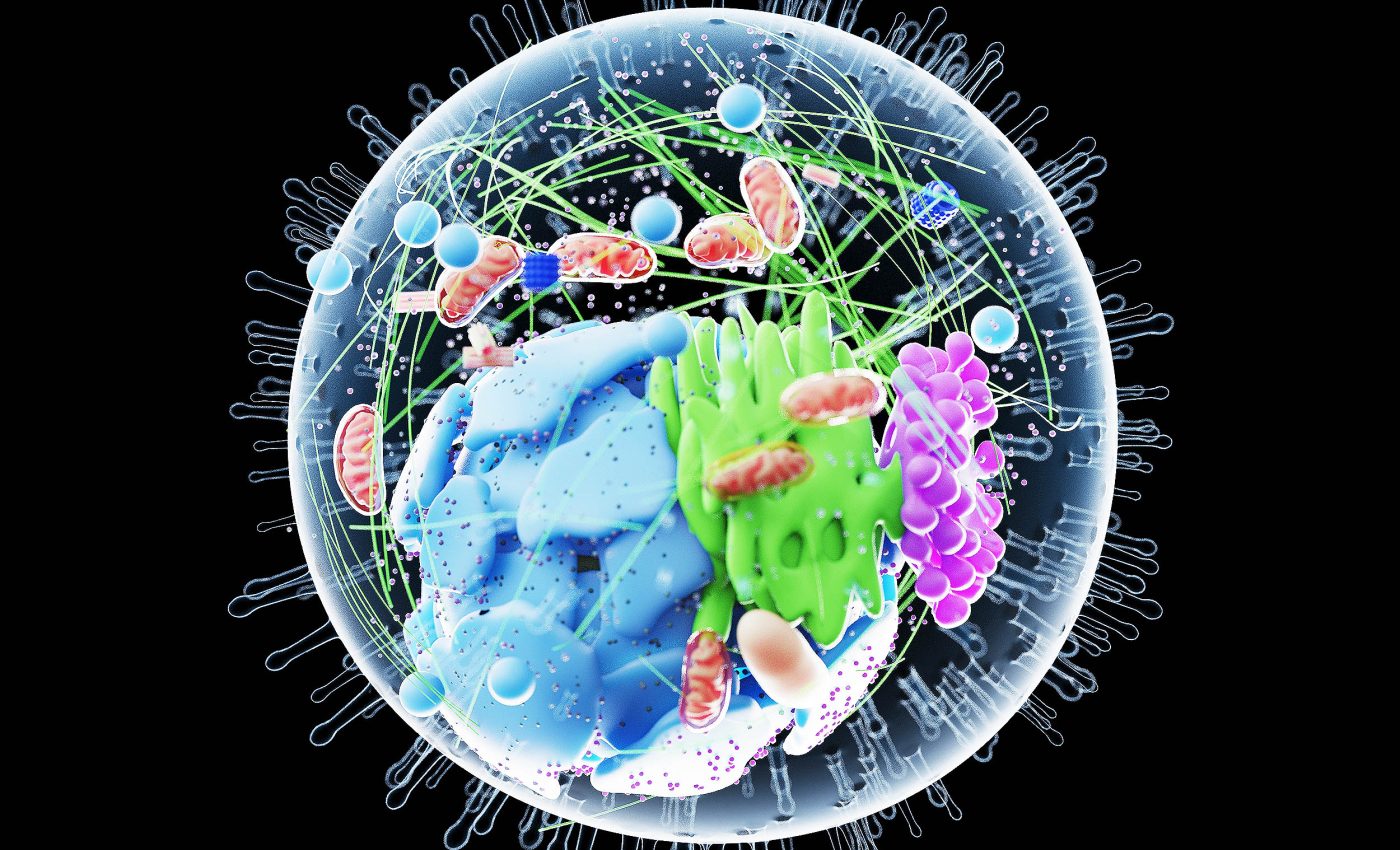
Cell biology is a world of constant motion and hidden structures. Much of what we know about cells comes from decades of research using microscopes, stains, and models. Yet, even in this well-charted territory, surprises still emerge – enter the “hemifusome.”
This previously unknown organelle may help explain how cells sort, recycle, and discard their internal cargo. This function is vital to life and is often disrupted in instances of genetic disease.

The discovery of this organelle, which was made by scientists at the University of Virginia and the National Institutes of Health (NIH), offers a new lens through which to study the inner workings of the cell.
It also presents a possible turning point for understanding diseases where cellular housekeeping breaks down. Using cutting-edge imaging tools, researchers have caught this organelle in action and outlined its potential impact on health and medicine.
Introducing the hemifusome
The hemifusome is not a static component but a temporary structure that appears and disappears depending on the cell’s needs.
It consists of two vesicles joined together by a partial membrane connection called a hemifusion diaphragm.
In this configuration, the vesicles do not fully merge but maintain a shared boundary that allows them to interact without blending entirely.
“This is like discovering a new recycling center inside the cell,” said researcher Seham Ebrahim, Ph.D., of UVA’s Department of Molecular Physiology and Biological Physics.
“We think the hemifusome helps manage how cells package and process material, and when this goes wrong, it may contribute to diseases that affect many systems in the body.”
These hemifused vesicles appear in two configurations. In the direct form, a smaller vesicle is attached to the outer side of a larger one, whereas in the flipped version, the smaller vesicle is embedded on the inner, or luminal, side.
In both cases, a dense particle called a proteolipid nanodroplet anchors the structure at the junction, possibly guiding its formation and stability.
How the hemifusome appears
To study hemifusomes, researchers turned to cryo-electron tomography (cryo-ET). This imaging method freezes cells rapidly, preserving them close to their natural state.
Unlike traditional electron microscopy, which can distort or destroy delicate structures, cryo-ET allows scientists to see cellular architecture as it truly exists.
By scanning the outer edges of four mammalian cell types, COS-7, HeLa, RAT-1, and NIH/3T3, the team identified hundreds of hemifusomes. These organelles made up nearly 10 percent of all membrane-bound vesicles in those regions.

Their consistency across cell types suggests they are not rare anomalies but common cellular components.
“You can think of vesicles like little delivery trucks inside the cell,” said Ebrahim, of UVA’s Center for Membrane and Cell Physiology. “The hemifusome is like a loading dock where they connect and transfer cargo. It’s a step in the process we didn’t know existed.”
What makes the hemifusome unique
Hemifusomes stand out not just for their shape, but for what’s inside them. The larger vesicle usually contains granular material, similar to what is seen in endosomes and ribosome-associated vesicles.
But the smaller vesicle shows a smooth, translucent interior. This likely reflects a protein-free or dilute aqueous solution, setting it apart from other vesicles in the cell.
The hemifusion diaphragm itself is unusually large, about 160 nanometers in diameter, far bigger than the 10 nanometer diaphragms seen in standard vesicle fusion events. These extended diaphragms appear stable, not fleeting, suggesting they may be designed to last.
In some cases, the diaphragm grows large enough to engulf the entire smaller vesicle into the larger one’s bilayer, creating a lens-like shape known in simulations as dead-end hemifusion. Seeing this in actual cells challenges the idea that such formations are purely theoretical.
Anchors and architects of the organelle
One consistent feature at the heart of hemifusomes is the dense proteolipid nanodroplet, or PND. About 42 nanometers in diameter, these droplets are lodged at the rim of the hemifusion site.
Their content, lipids and proteins, suggests they may help build or stabilize the hemifused structure.
These PNDs have never been observed in such a role before. Some appear free in the cytoplasm, others are embedded in membranes. Researchers propose that PNDs may serve as scaffolds for assembling new vesicles.
As the PND integrates into a membrane, it may kickstart the formation of the smaller vesicle seen in hemifusomes.
This process, described as de novo vesiculogenesis, stands apart from classical vesicle fusion. The presence of a unique, translucent vesicle and the absence of known docking steps indicate the hemifusome may follow its own assembly path.
Are hemifusomes related to endosomes?
Given their location and size, hemifusomes resemble some endosomal structures. To investigate this further, the researchers traced the journey of gold nanoparticles, common markers used to map endocytic activity.
The particles entered known endosomes and lysosomes but never appeared inside hemifusomes. This absence suggests that hemifusomes do not belong to the classical endocytic pathway.
Instead, they may represent a separate system operating independently of the cargo sorting carried out by proteins like ESCRT. This distinction may have wide implications for how we understand vesicle traffic inside cells.
Multivesicular bodies and disease
Some hemifusomes evolve into more complex structures. The study observed compound hemifusomes that contained multiple vesicles, all partially fused.
These could be early versions of multivesicular bodies (MVBs), which cells use to break down and recycle internal material.
In the canonical model, ESCRT proteins form inward buds that eventually pinch off inside a larger vesicle. But in hemifusomes, vesicles grow inward through hemifusion and expand with the help of PNDs.
This alternative route might explain how MVBs form in ways not covered by traditional theories.
One such condition affected by these pathways is Hermansky-Pudlak syndrome. It is a genetic disorder marked by defects in pigmentation, lung function, vision, and bleeding. Cellular recycling issues are central to the disease.
Understanding the hemifusome may help explain these disruptions and lead to future treatments.
A new model for vesicle formation
The study proposes a full model where PNDs trigger the formation of translucent vesicles that partially fuse with larger ones, forming hemifusomes.
These structures may then bud inward, transforming into flipped hemifusomes. Over time, they could scission off as free vesicles inside MVBs.
In contrast to the ESCRT system, which requires tight protein coordination, this mechanism relies on structural and biophysical cues.
It also sidesteps the need for large lipid donations from other organelles, solving a long-standing puzzle in vesicle formation research.
“This is just the beginning,” Ebrahim said. “Now that we know hemifusomes exist, we can start asking how they behave in healthy cells and what happens when things go wrong. That could lead us to new strategies for treating complex genetic diseases.”
What comes next
The implications of this discovery stretch far beyond cell biology. By offering a new pathway for how cells build and manage internal compartments, the hemifusome challenges decades of assumptions.
It also invites new thinking about disease, especially conditions where cells fail to manage their waste.
Future research will focus on identifying what proteins guide hemifusome formation and how PNDs are created.
Scientists also want to know if these structures exist in other parts of the cell, not just at the periphery. Advanced imaging tools and genetic models will be key to answering these questions.
In a field where many believed the major organelles were already mapped, the hemifusome serves as a reminder. The cell still holds secrets. And some of them could lead to cures.
The study is published in the journal Nature Communications.
—–
Like what you read? Subscribe to our newsletter for engaging articles, exclusive content, and the latest updates.
Check us out on EarthSnap, a free app brought to you by Eric Ralls and Earth.com.
—–


