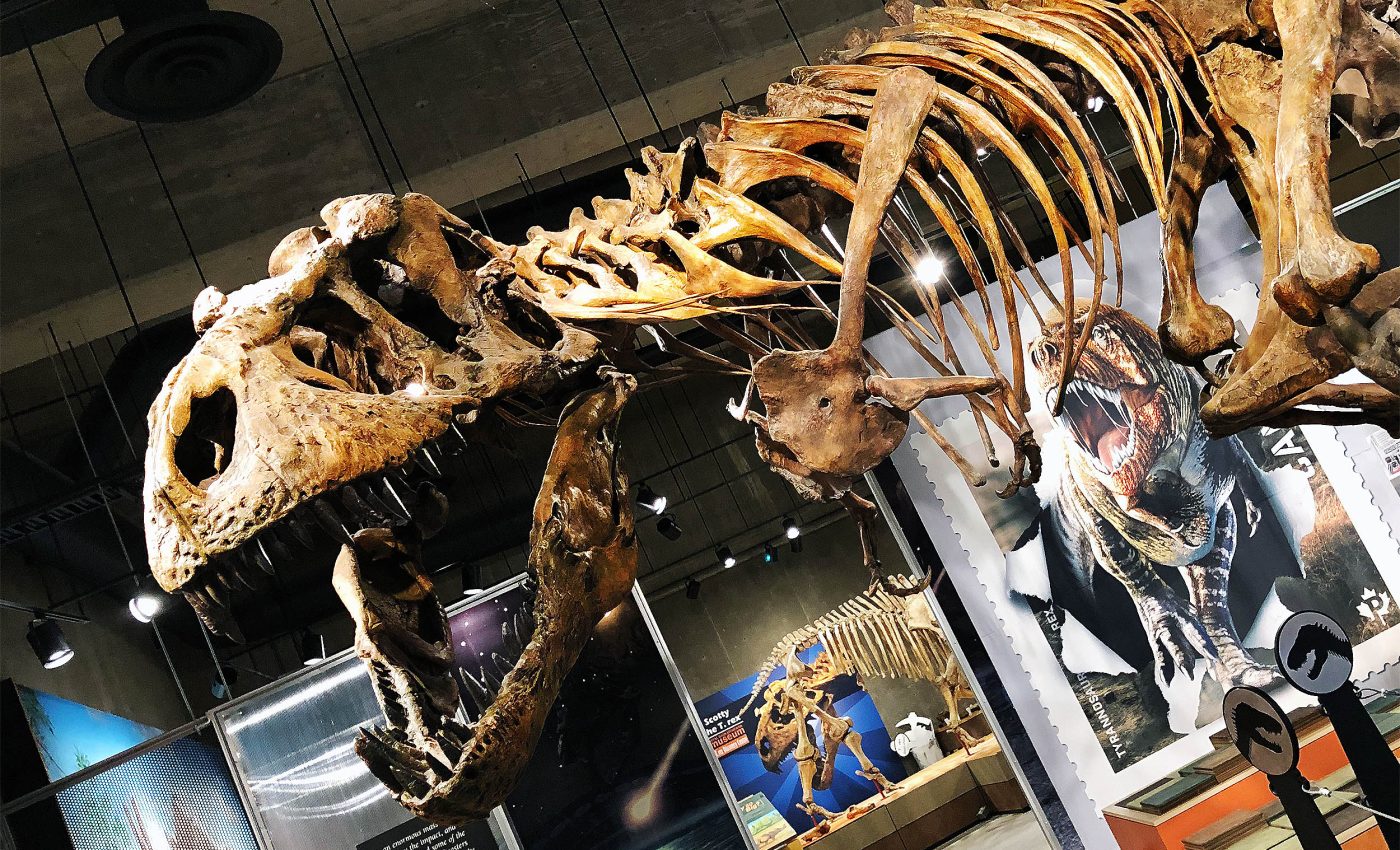
Preserved blood vessels found in bones of the world's largest T. rex
A broken bone sets off a repair job inside the body. New blood vessels grow in, bringing oxygen and nutrients to rebuild the damage.
Scientists have now traced that same process in a Tyrannosaurus rex rib from about 67 million years ago, using powerful X-rays to see mineral “shadows” where blood once flowed.

They focused on a deep fracture near the vertebral end of the rib. The bone had started to heal before the animal died, laying down a callus – the bumpy repair tissue that forms during recovery.
Inside that callus, they spotted tube-like structures that matched the size and pattern of blood vessels linked with healing.
Finding T. rex blood vessels
The work centers on a Tyrannosaurus rex specimen named “Scotty,” housed at the Royal Saskatchewan Museum in Canada, one of the largest and most complete T. rex skeletons ever found.
The research team scanned the rib at the Canadian Light Source synchrotron, a facility that generates extremely bright X-rays for high-resolution imaging and chemical analysis.
Jerit L. Mitchell, a PhD student in the Department of Physics at the University of Regina, led the study after joining the project during the first scan of the rib in 2019.
“I remember showing my supervisors, Dr. Barbi and Dr. McKellar, a strange structure inside a scan of the rib that I originally didn’t give much thought to,” Mitchell reminisced.
“They were quick to point out that what I discovered could possibly be preserved blood vessels, which has since led to a much more expansive research project.”
Blood vessels from an injured T. rex
The vessel-like structures only showed up around the fracture and its callus, not throughout the rib. They were larger than the tiny Haversian canals that normally carry blood in compact bone.
That pattern lines up with what doctors see today: during healing, the body ramps up angiogenesis – growth of new vessels – to flood the injured area with supplies.
The team kept the features in place, studying them “in situ” rather than grinding the fossil down. That choice protected a rare specimen and let the researchers compare 3D shapes and chemistry directly within the bone.
High-tech tools
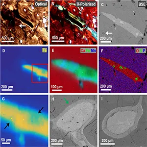
Synchrotron micro-CT produced 3D images at micrometer-scale resolution, small enough to resolve fine tubes weaving through the callus.
X-ray fluorescence (XRF) mapped elements, and X-ray absorption near-edge structure (XANES) identified their chemical states.
The team also checked thin sections with light and electron microscopes to confirm textures and mineral identities.
Together, those methods allowed them to connect geometry with chemistry. They could tell not just that tubes were present, but also what filled them and how those fillings changed over time.
Iron, fractures, and soft tissue
The tubes were rich in pyrite, an iron sulfide often called fool’s gold. Some of that pyrite later oxidized to iron oxides such as goethite and hematite.
Iron can help stabilize organic structures during fossilization, and the resulting minerals can persist long after soft tissue disappears.
Here, the original vessels are gone, but their mineral casts remain, recording the paths where blood once moved through healing bone.
“Normally, what gets preserved in the fossil record is only just the hard parts – just the bones or the teeth,” Mitchell told CBC News’ Adam Hunter.
“But we can actually have the soft tissues preserved in rare circumstances, and these can tell us a lot more about how dinosaurs lived millions of years ago.”
The chemistry and the location work together. Iron sulfides transitioning to iron oxides fit a plausible path from soft tissue to mineral replicas, especially in a confined space like a fracture callus where blood flow once surged.
Location of the T. rex blood vessels
The features cluster where healing was most active. Temporary vessels can grow larger than everyday bone canals as they branch from the marrow cavity and the bone surface to supply repair tissue.
Seeing that size and distribution in the rib strengthens the case that these were angiogenesis-related structures, not ordinary anatomy or random mineral growth.
“Preserved blood vessel structures, like we have found in Scotty’s rib bone, appear linked to areas where the bone was healing. This is because during the healing process, those areas had increased blood flow to them,” Barbi explained.
This targeted preservation also speaks to how fossils can capture brief, intense biological episodes. A break triggers a rush of activity lasting weeks or months.
Under the right burial conditions, traces of that activity can harden into rock and wait for us to find them.
“This work also provides a new way to compare how injuries healed in extinct animals, like dinosaurs, with living species, such as birds and reptiles, which helps us better understand the biology of the past, and also how life on Earth has evolved over millions of years,” Barbi concluded.
Why does any of this matter?
These results connect dinosaur biology with processes we see in clinics and labs today.
Bone is living tissue. It remodels, it repairs, and – under certain conditions – its repair work can leave a chemical and structural record that lasts.
The study also shows how non-destructive tools can reveal fragile features without harming invaluable fossils.
The approach helps clarify past debates over “soft tissues” in dinosaur bones. Here, the authors do not claim original vessels.
They present mineral casts that track vessel paths in a healing zone, supported by size, distribution, and iron-based chemistry.
Lessons learned for future studies
Healed injuries may be prime places to look for similar traces in other dinosaurs and in distant relatives like early birds and reptiles.
With broader sampling, finer imaging, and detailed chemical work, researchers could compare healing rates, test links to metabolism, and study how burial environments shape preservation.
A single broken rib from an enormous T. rex skeleton has told a very precise story: blood rushed to a wound, vessels expanded, bone knit, and minerals later filled the spaces left behind.
The biology ended ages ago. The imprint stayed put.
The full study was published in the journal Scientific Reports.
—–
Like what you read? Subscribe to our newsletter for engaging articles, exclusive content, and the latest updates.
Check us out on EarthSnap, a free app brought to you by Eric Ralls and Earth.com.
—–


