Oculomics: How AI is turning eye scans into full-body health checks and disease detection
The core idea behind a rapidly-emerging field of science called oculomics is simple and bold: detailed eye exams may reveal far more about overall human health than just how well a person can see.
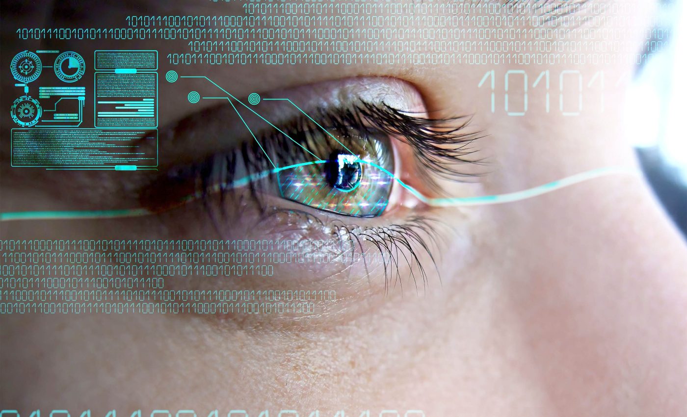
High-resolution retinal images, paired with modern AI algorithms, are now being used to detect risks for diseases that reach far beyond eyesight.

Large-scale datasets are already fueling this revolution. The UK Biobank, for instance, houses retinal and health data from more than 500,000 volunteers, giving scientists an unprecedented window into how subtle eye features mirror the body’s overall health.
Understanding oculomics – the basics
Cutting-edge technology used in the nascent field of oculomics allows scientists and doctors to uncover signs of heart disease, diabetes, kidney issues, and even neurological disorders – all from a quick, noninvasive eye scan.
The human retina is an extension of the brain and shares blood vessels with other vital organs. This means tiny changes in the retina’s structure or color can signal bigger problems elsewhere in the body, long before symptoms appear elsewhere.
Advanced AI algorithms analyze high-resolution images of the retina, optic nerve, and blood vessels to detect patterns far too subtle for the human eye.
These AI systems can measure vessel width, nerve fiber thickness, pigmentation, and even microstructural changes in the retina.
By combining ocular data with genetic, lifestyle, and clinical information, AI used in oculomics can create what researchers call a “digital eyeprint” – a personal health signature that reflects how well your body is functioning as a whole.
Retinal link to systemic disease
The work was led by Dr. Tien Yin Wong at the Singapore National Eye Centre and Duke NUS Medical School (SNEC). His research centers on retinal imaging, artificial intelligence, and how ocular clues relate to systemic disease.
The retina, a thin light-sensing layer at the back of the eye, exposes tiny blood vessels and nerve tissue with uncommon clarity. Those are two organ systems that usually cannot be examined directly without surgery.
Oculomics connects small changes in retinal structure and blood flow to patterns seen in the heart, brain, and kidneys.
Oculomics and AI
A deep-learning study trained on 284,335 patients showed that retinal photographs can estimate common risk factors such as age and smoking status.
It also predicted the likelihood of a major cardiac event within five years – with performance comparable to standard calculators.
Deep learning, a machine-learning method that identifies patterns from large datasets, excels at finding subtle image features that humans miss. It weighs vessel width, twists, and textures, then converts them into a composite risk score.
Health clues in every eye photo
A multi-country system used retinal photos to estimate coronary artery calcium – calcium buildup in heart arteries that indicates plaque burden – and stratified cardiovascular risk without a CT scan.
The approach offered a low-cost, noninvasive way to identify who might need more intensive testing.
A separate algorithm detected Alzheimer’s disease from retinal photographs in a multicenter sample. The model captured disease-linked features in the macula and microvasculature while standardizing results across sites.
Another team reported a retinal age-gap biomarker that correlated with mortality risk. When the predicted retinal age exceeded a person’s actual age, risk increased modestly but consistently.
AI joins primary care
“Today’s decision permits the marketing of a novel artificial intelligence technology that can be used in a primary care doctor’s office,” said Dr. Malvina Eydelman, an FDA device director.
Regulators have started to permit autonomous tools that interpret retinal images in primary care.
That early green light focused on diabetic retinopathy, but it set a clear precedent. Autonomous AI, software that can make a clinical call without human oversight, could triage patients where specialists are scarce.
Oculomics still has limitations
The clearest successes so far lie in high-burden conditions with well-defined biological signatures in the eye. Heart risk, cognitive decline, diabetes, and kidney stress all leave detectable traces in vessel shape or retinal layers.
Limits still matter. Generalizability – the ability of a model to perform reliably across new clinics and populations – can falter when cameras, image quality, or patient backgrounds differ from the training data.
Clinicians should treat oculomics outputs like any other biomarker, a measurable signal that reflects disease risk. A positive result may prompt follow-up tests, not an instant diagnosis.
False alarms and misses will happen, especially at the edges. That is why teams are validating models prospectively, recalibrating thresholds, and combining image signals with routine vitals.
Eyes as health sensors
The microvasculature – the body’s smallest blood vessels that carry oxygen – is visible in every retinal image, offering a clear snapshot of vascular health throughout the body.
Modern cameras now make that window easy to access: a 30-second, noninvasive photo can be taken in a primary care clinic, a pharmacy, or even by mobile teams during community screenings.
Portable imaging systems and home-based vision tools are extending that reach even further.
As datasets expand, foundation models – large pre-trained AI systems that can adapt to many tasks – are expected to improve stability and reduce the need for manual labeling.
Ultimately, the goal is precision prevention. By translating subtle eye signals into actionable health insights, oculomics could help match each patient to the right “next step,” whether that means starting a statin, investigating cognitive decline, or tightening glucose control.
The study is published in Progress in Retinal and Eye Research.
—–
Like what you read? Subscribe to our newsletter for engaging articles, exclusive content, and the latest updates.
Check us out on EarthSnap, a free app brought to you by Eric Ralls and Earth.com.
—–


